Call Us Today!
(718) 347-6262

Digital radiography has transformed the way dental imaging supports diagnosis and treatment. By replacing traditional film with electronic sensors and computer processing, modern dental practices can capture high-resolution images in seconds and use them immediately to guide care. For patients, that means quicker visits, clearer explanations from your provider, and a record that integrates seamlessly with your digital chart.
At its core, digital radiography uses a compact sensor placed in the mouth or an external receptor to capture X-ray images. Those sensors convert X-ray energy into digital signals that a computer processes into detailed images. Because the process eliminates film development, patients no longer wait for physical processing; images appear on-screen almost instantly.
One of the most tangible benefits for patients is clarity. Digital images can be enlarged, enhanced, and adjusted for contrast so clinicians can examine fine details that might be harder to see on film. That improved visibility supports earlier detection of cavities, bone changes, and other oral-health concerns that benefit from prompt attention.
Another practical advantage is convenience. While the technical steps happen behind the scenes, patients experience shorter chair time and the ability to view their images with the dentist. Seeing an image alongside an explanation helps patients understand their oral health and participate more confidently in treatment planning.
Digital imaging delivers higher-resolution results than traditional film in many cases, and the software tools available with these systems amplify diagnostic potential. Clinicians can zoom in on suspicious areas, adjust brightness and contrast, and compare current images to previous records, which supports more accurate assessments over time.
Because digital images are available immediately, treatment decisions are no longer delayed by film processing. This speed can be especially important for urgent situations where rapid evaluation is necessary. Multiple members of the dental team can review images at the same time, facilitating collaborative decision-making and coordinated care.
Image manipulation is done without altering the original file, preserving the integrity of the record while allowing clinicians to highlight specific findings for patients. This combination of speed, flexibility, and preservation of source data strengthens clinical confidence when creating treatment plans.
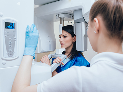
When a radiograph is needed, a small sensor or detector is positioned carefully to capture the area of interest. The equipment is designed with patient comfort in mind, and sensors are lightweight and thin to minimize gag reflex and reduce discomfort. Once the image is taken, it is transmitted directly to the computer and associated with the patient’s electronic record.
In practice, this means less time spent waiting for images to develop and more time focused on explaining findings and discussing next steps. The immediate availability of images helps the dental team answer questions on the spot and create an efficient plan for any recommended care, whether preventive or restorative.
To ensure consistent image quality, sensors and software are maintained and calibrated according to best-practice protocols. Patients benefit from a system that is not only faster but also subject to routine quality checks so clinicians can rely on accurate, repeatable results.
Because digital radiographs become part of the electronic health record, they simplify long-term recordkeeping and follow-up. Images can be archived in a patient’s chart, retrieved quickly for comparison, and used to document progression or healing after treatment. That continuity is valuable for monitoring chronic conditions and evaluating outcomes over time.
Digital files can also be shared securely with specialists when collaborative care is needed. Whether coordinating with an endodontist, oral surgeon, or orthodontist, digital radiography allows for fast transfer of detailed images so consultations can proceed without delay. This connected workflow improves coordination and helps ensure patients receive comprehensive care.
For patient education, digital images are a powerful tool. Dentists can display and annotate images on-screen to illustrate a diagnosis or show why a recommended treatment is appropriate. Visual explanations often make complex topics more accessible, enabling patients to make informed choices about their care.
Digital radiography reduces radiation exposure compared with many conventional film systems because modern sensors are more efficient at capturing image data. Clinicians still adhere to established safety protocols — using protective aprons, limiting the number of images to those necessary for diagnosis, and following ALARA (as low as reasonably achievable) principles — to minimize exposure for every patient.
From an environmental standpoint, eliminating film development removes the need for chemical processing and paper waste associated with traditional X-rays. That reduction in consumables makes digital systems a more eco-conscious choice for practices aiming to lower their environmental footprint while maintaining high standards of care.
Comfort and trust go hand-in-hand: patients benefit from quicker procedures and fewer repeat images thanks to advanced sensors and imaging workflows. When combined with routine maintenance and safety checks, digital radiography contributes to a patient-centered experience that prioritizes clarity, efficiency, and well-being.
At the office of David M. Goldberg, DDS, digital radiography is part of a broader commitment to providing precise, contemporary dental care. Our team uses these tools to support accurate diagnoses, clear communication, and coordinated treatment plans that put patients’ needs first.
In summary, digital radiography offers faster imaging, improved diagnostic detail, safer exposure levels, and streamlined recordkeeping. If you have questions about how digital X-rays are used in our practice or what to expect during your visit, please contact us for more information.
Digital radiography captures dental images using electronic sensors and computer processing instead of chemical film. The sensors convert X-ray energy into digital files that appear on-screen within seconds, eliminating development time and physical film handling. This immediate availability speeds diagnosis and allows clinicians to manipulate images for clearer viewing.
The resulting files can be enlarged, adjusted for contrast, and compared side-by-side with prior images without degrading the original record. Because the process is electronic, images integrate directly into the patient’s digital chart, simplifying recordkeeping and long-term monitoring. These workflow improvements are a primary reason many practices have moved from film to digital systems.
Digital images typically offer higher resolution and sharper contrast than conventional film, and software tools enhance the clinician’s ability to detect fine details. Dentists can zoom in on suspicious areas, adjust brightness and contrast, and use measurement tools to evaluate bone levels or root anatomy more precisely. These capabilities support earlier detection of cavities, bone changes, and other conditions that benefit from prompt attention.
Immediate access to images also streamlines clinical decision-making by allowing the dental team to review findings during the same visit and discuss options with patients. Comparing current images to archived records helps track progression or healing over time and supports evidence-based treatment planning. Overall, better visualization and faster workflows contribute to more accurate, coordinated care.
Digital radiography generally exposes patients to lower doses of radiation than many traditional film systems because modern sensors are more efficient at capturing image data. Clinicians continue to follow established safety practices, including the use of protective aprons, thyroid collars when appropriate, and the ALARA principle to keep exposure as low as reasonably achievable. Only images necessary for diagnosis are taken, which further reduces unnecessary exposure.
Pregnant patients and those with specific medical concerns should always inform their dentist so the team can take appropriate precautions or defer imaging when clinically acceptable. Quality control, proper shielding, and trained operators together ensure that digital radiography remains a safe and effective diagnostic tool. If you have specific concerns about radiation, your dentist can explain the measures used in the practice to protect you.
When a radiograph is needed at the office of David M. Goldberg, DDS, a small intraoral sensor or external receptor is positioned to capture the area of interest, and the image is taken in seconds. Sensors are lightweight and thin to minimize discomfort and are positioned carefully to limit gag reflex and ensure accurate images. The captured image is transmitted instantly to the computer and associated with the patient’s electronic record for review.
Because images appear immediately, the dental team can review findings with the patient during the same visit, answer questions, and discuss next steps without delay. Routine maintenance and calibration of sensors and software are performed to preserve image quality and reliability. This combination of comfort, speed, and integration supports efficient, patient-centered care.
Preparation for a dental X-ray appointment is minimal; patients should arrive with any recent dental records or images if they were taken elsewhere. You should inform the dental team about pregnancy, recent medical imaging, and any implants or devices in the mouth that may affect imaging. Wearing comfortable clothing and removing necklaces or glasses that could interfere with head positioning will also help streamline the process.
The dental team will explain the procedure before taking images and will provide protective shielding as needed, such as a lead apron or thyroid collar. If you have concerns about comfort or anxiety, let the staff know so they can adjust positioning or pacing. Clear communication ensures the visit is efficient and comfortable for you.
The frequency of dental radiographs depends on individual risk factors, oral health status, and the specific clinical questions the dentist needs to answer. Routine schedules vary: some patients with excellent oral health and low risk may need images less often, while those with active disease, ongoing treatment, or certain systemic conditions may require more frequent imaging. Your dentist will recommend a schedule tailored to your needs based on clinical examination and history.
Because digital radiographs are readily available for comparison, they are valuable for monitoring changes over time and documenting healing after treatment. Rather than following a one-size-fits-all timeline, the practice uses clinical judgment and evidence-based guidelines to determine when images are necessary. Discuss your individual risk and the rationale for recommended imaging with your provider to understand the plan for your care.
Yes. One of the practical advantages of digital radiography is the ability to securely share high-quality images with specialists, other providers, or referring clinicians. Digital files can be exported or transferred through secure channels, enabling timely consultations with endodontists, oral surgeons, orthodontists, or other specialists without the delays associated with physical film. This capability improves coordination and helps streamline multi-disciplinary care.
Digital images are also archived in the patient’s electronic chart for future comparison and long-term monitoring. Having readily retrievable images simplifies follow-up care and supports evidence-based decisions when evaluating progression or healing. Proper data management and secure transfer protocols protect patient privacy while allowing efficient collaboration.
Digital radiography is well suited for pediatric and adolescent patients because modern sensors capture quality images quickly, which reduces time in the chair and the likelihood of repeat exposures. Lower radiation doses and the ability to take fewer, targeted images make digital systems advantageous for young patients. Sensors designed for smaller mouths improve comfort and positioning for more reliable results.
Immediate on-screen viewing also helps clinicians explain findings to parents and adolescents using visual aids, which can improve understanding and cooperation. When combined with age-appropriate explanations and behavioral techniques, digital imaging supports efficient, child-friendly care. The practice tailors imaging protocols to the child’s developmental stage and clinical needs to ensure both safety and diagnostic value.
Maintaining reliable imaging requires routine calibration of sensors, software updates, and scheduled equipment checks to ensure consistent performance. The practice follows manufacturer recommendations and clinical best-practice protocols for cleaning, inspection, and calibration so that images remain accurate and reproducible. Regular staff training in positioning and exposure technique also reduces the need for repeat images and maintains diagnostic quality.
Recordkeeping of maintenance activities and periodic review of image quality are part of a structured quality assurance program that supports dependable results. When equipment anomalies are identified, prompt servicing or replacement prevents compromised imaging. These measures help clinicians rely on digital radiographs for clear, actionable diagnostic information.
The practice uses digital radiographs as a visual tool to explain diagnoses and illustrate why a recommended treatment is appropriate, which helps patients make informed decisions. Dentists can annotate images, highlight areas of concern, and compare current and past images to show progression or healing, making complex information more accessible. This visual approach supports shared decision-making and increases patient confidence in the proposed plan.
Because images are integrated into the electronic record, they can be revisited during follow-up visits to show results and document outcomes. Clear communication combined with immediate visual evidence strengthens the clinician–patient relationship and ensures that recommendations are transparent and evidence based. If you have questions about findings or options, the team will review the images with you in detail.